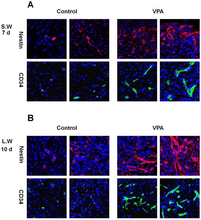
Figure 5. Effects of VPA on the expression of stem cell markers in wounds.
A full-thickness skin excision (diameter = 0.5 cm or 1.5 cm) was made on the backs of 8-week-old C3H mice, and 500 mM VPA was topically applied to the wounds daily. (A) Immunohistochemical analysis of Nestin or CD34 in the control and VPA-treated small wounds (original magnification ×635). (B) Immunohistochemical analysis of Nestin or CD34 in the control and VPA-treated large wounds (original magnification ×635).

