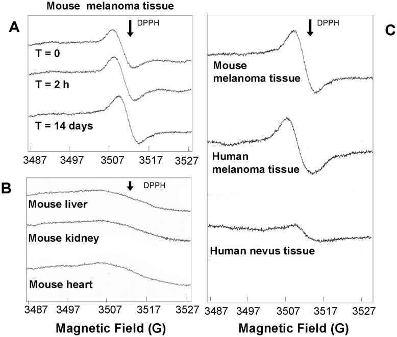
Figure 2. ESR spectra of murine- and human- melanoma and healthy tissues.
A) Murine B16F10 melanoma cells were injected in 5 mice in order to produce primary melanomas. Mice were sacrificed 14 days after the cell injection and tumours were collected for ESR analysis. The spectra show the presence of a strong signal located at the same position as observed in human melanoma cells. Signal was stable over time (recorded after 2 hours and after 14 days upon frozen storage). B) Murine tissues from liver, kidney and heart do not show ESR signal in the same magnetic field range. C) ESR spectra of formalin-fixed paraffin-embedded sections of human melanoma, human nevus tissue and fresh mouse melanoma tissue. DPPH arrow indicates the position of the standard free radical signal (1, 1-diphenyl-2-picrylhydrazyl).

