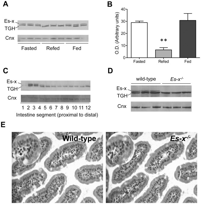
Figure 1. Intestinal Ces1/Es-x expression is regulated by nutritional status.
(A) Intestinal Ces1/Es-x protein expression in different nutritional states. Mice were fasted for 24 h and refed for 6 h. Fasted and refed mice were euthanized at 8:00 P.M. TGH is a related carboxylesterase migrating at a lower Mr due to lesser glycosylation and is recognized by the polyclonal anti-Ces1/Es-x antibodies. Two µg intestinal proteins were subjected to analysis. Cnx, calnexin = loading control. (B) Quantitation of Ces1/Es-x immunoreactive bands obtained in different nutritional states. **p<0.01 Refed vs Fasted. (C) Ces1/Es-x protein distribution along the small intestine. Small intestine was cut into 12 pieces 2-cm long. Proteins were separated by SDS-PAGE and immunodetected with anti-Ces1/Es-x antibodies. Representative data from 3 different independent experiments are shown. (D) Absence of Ces1/Es-x protein (immunoblot) in the intestine from Ces1/Es-x −/− mice. (E) H&E staining of small intestine (200×) sections.

