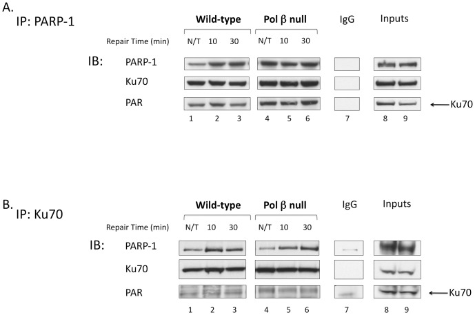
Figure 5. Interaction of PARP-1 and Ku70 after MMS exposure.
A. Immunoprecipitation with anti-PARP-1 antibody from untreated (N/T) and MMS-treated cell extracts with the repair times specified. B. Immunoprecipitation with anti-Ku70 antibody as in A. Immunoblotting (IB) was performed with the antibodies specified. An IgG negative control was performed for both immunoprecipitations (lanes 7), and 30 µg of cell extract (input wild-type and pol β null, respectively) was used as a source of marker proteins (lanes 8 and 9).

