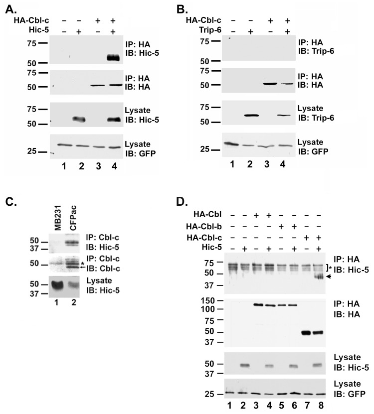
Figure 1. Hic-5 interacts with Cbl-c.
A. HEK293T cells were transfected with HA-Cbl-c alone, Hic-5, or the combination of HA-Cbl-c and Hic-5 as indicated above panels. B. HEK293T cells were transfected with HA-Cbl-c alone, Trip-6, or the combination of HA-Cbl-c and Trip-6 as indicated above panels. In A and B, HA-Cbl-c was immunoprecipitated from the cell lysates. Immunoprecipitates (IP) or cell lysates (lysate) were immunoblotted (IB) as indicated to the right of the panels. All transfections were balanced with empty vector controls. Green fluorescent protein (GFP) was transfected as a control for transfection efficiency and is shown as a loading control. C. Endogenous Cbl-c was immunoprecipitated from whole cell lysates from MB231 (which expresses Hic-5 but no Cbl-c) and CFPAC-1 (which expresses both Hic-5 and Cbl-c) cell lines and immunoblotted for Cbl-c and Hic-5. Immunoprecipitates (IP) or cell lysates (lysate) were immunoblotted (IB) as indicated to the right of the panels. The arrow indicates immunoprecipitated Cbl-c and the asterisk indicates the immunoglobulin heavy chain. D. HEK293T cells were transfected with HA-Cbl, HA-Cbl-b, HA-Cbl-c alone, Hic-5 alone, or each HA epitope tagged Cbl protein with Hic-5 as indicated above the panels. Cbl proteins were immunoprecipitated and immunoprecipitates (IP) or cell lysates (lysate) were immunoblotted (IB) as indicated to the right of the panels. The arrow indicates immunoprecipitated Hic-5 and the asterisk indicates an artifact band that includes the immunoglobulin heavy chain. MWs in kDa are shown to the left of the panels.

