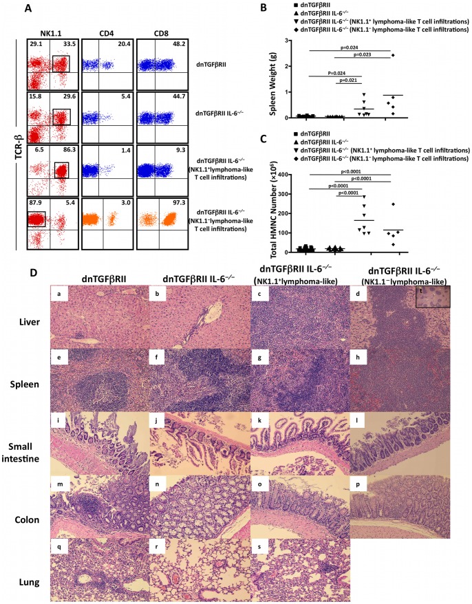
Figure 2. Histological features and immunophenotypes of lymphoma-like T cell infiltration.
A. Flow cytometric analysis of HMNCs from dnTGFβRII and dnTGFβRII IL-6−/− mice with and without lymphatomous lesion. The numbers above the plots indicate the frequency of TCRβ+NK1.1− and TCRβ+NK1.1+ cells (left panels), the frequency of CD4 positive cells (middle panels) and the frequency of CD8 positive cells (right panels). Cells shown in the middle and right panels were gated on TCRβ+NK1.1− or TCRβ+NK1.1+ populations as indicated in the left panels. B. The spleen weight of dnTGFβRII, dnTGFβRII IL-6−/− and dnTGFβRII IL-6−/− mice with a predominant NK1.1 positive or negative phenotype at age of 24–40 weeks. C. The total HMNC counts of dnTGFβRII, dnTGFβRII IL-6−/− and dnTGFβRII IL-6−/− mice with a predominant NK1.1 positive or negative phenotype at age of 24–40 weeks. D. Representative H&E stained sections of tissue sample including liver (a–d), spleen (e–h), small intestine (i–l), colon (m–p) and lung (q–s) were prepared from dnTGFβRII and dnTGFβRII IL-6−/− mice at age of 24–40 weeks (a–s,×200; t,×40). Typical diffuse lymphomatous lesions were found in liver (c) and spleen (g) of dnTGFβRII IL-6−/− mice with a predominant NK1.1+ phenotype, while large focal lymphomatous lesions were found in liver (d,×200)(t,×40) and spleen (h) of dnTGFβRII IL-6−/− mice with a predominant TCRβ+ NK1.1− phenotype. No obvious lymphomatous lesions were found in lung, small intestine and colon in dnTGFβRII and dnTGFβRII IL-6−/− mice.

