
Figure 2. Structures and properties of geldanamycin derivatives.
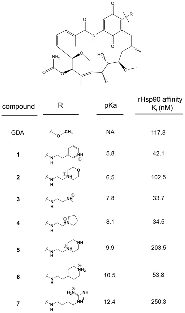
The seven derivatives with different weak base modifications at the 17-position of GDA are shown. The pKa values for derivatives 1, 2, 3, 5 and 6 was measured experimentally using proton NMR (n = 1). The pKa for compound 4 was measured experimentally in an earlier manuscript (see Results). The pKa for compound 7 was estimated using software (see Materials and Methods). The binding affinity for each of the inhibitors with rHsp90 is shown (n = 2), and was based on a previously established fluorescence polarization assay (see Materials and Methods).