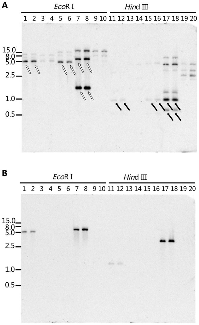
Figure 2. Identification of the Cyt b gene intron in Monilinia spp.
Two restriction enzymes EcoR I and Hind III were used to generate the restriction profiles. Digested genomic DNA was separated in 0.8% agarose gel, and the blot was hybridized with the My3 (A) and My4 (B) fragments. White and black arrows in (A) indicate the expected bands for EcoR I and Hind III digestions. Lanes 1, 11 and 2, 12: M. yunnanensis isolates YKG10-64a and SM09-7c; 3, 13 and 4, 14: M. fructigena isolates SL10 and Mfg2-GE-A; 5, 15 and 6, 16: M. laxa isolates BEK-SZ and EBR ba11b; 7, 17 and 8, 18: M. mumecola isolates HWL10-11a and HWL10-20a; 9, 19 and 10, 20: M. fructicola isolates MPA14 and BM09-4a. The sizes (in kilobases) of marker DNA fragments are indicated on both sides (Wide Range DNA Marker on the left and DL 2000 DNA Marker on the right, TaKaRa Biotechnology (Dalin) Co., Ltd).

