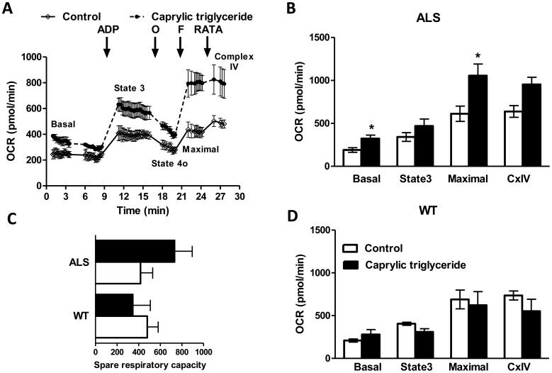
Figure 5. Mitochondrial bioenergetic profile in the spinal cord of WT and SOD1 G93A mice on control or caprylic triglyceride diet.
Mitochondria were isolated by differential centrifugation from the whole spinal cord of SOD1-G93A mice on control or caprylic triglyceride diet and oxygen consumption rates were analyzed using Seahorse XF24 extracellular flux analyzer. (A) A representative trace of OCR in the presence of pyruvate and malate. Adenosine diphosphate (ADP), oligomycin (O), carbonyl cyanide 4-(trifluoromethoxy)phenylhydrazone (FCCP) and a mixture of rotenone, antimycin A, N,N,N’,N’-tetramethylphenylenediamine and ascorbate (RATA) were injected at the indicated time points to measure basal, state 3, state 4o, maximal and complex IV OCR as indicated. OCR in the presence of pyruvate and malate in (B) SOD1-G93A (ALS) and (D) wild type (WT) mice. (C) Spare respiratory capacity of mitochondria from WT and ALS mice on control or caprylic triglyceride diet. Data are mean ± SEM, n = 3 for all groups, *p<0.05 as compared to control by two-tailed student t-test.

