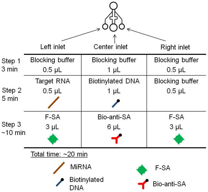
Figure 2. Schematic representation of the experimental protocol.
The center channels were always used for one reagent at a time. Step 1: 0.5 µL of blocking buffer (BB) was injected from all the inlets and incubated for 3 min. Step 2: 0.5 µL of the target miRNA sustained in BB, 1 µL of biotinylated probe DNA (10 nM in the BB), and 0.5 µL of BB, were injected from the left, center, and right inlets, respectively. Step 3: After 5 min of incubation, 3 µL of F-SA (2.5 µg/mL in BB), 6 µL of B-anti-SA (25 µg/mL in BB), and 3 µL of F-SA (2.5 µg/mL in BB) were injected from the left, center, and right inlets, respectively.

