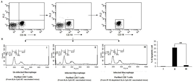
Figure 6. CD8+ T cell cytotoxicity. CD8+ T cells purified from SLA-CpG-DCs vaccinated parasitized mice 28 days after infection were co-incubated with autologous uninfected (I) and Leishmania-infected macrophages (II) in a 10∶1 ratio.
In another set, CD8+ T cells were purified from CXCL10 depleted SLA-CpG-DCs vaccinated parasitized mice 28 days after infection and co-incubated with autologous Leishmania-infected macrophages in a 10∶1 ratio (III). After 4 h, the cells were harvested and stained with anti-CD14-FITC antibodies to select the target macrophages (A). The dot plots were derived from the gated events based on the region encircling positive cells. This CD14-FITC positive population were analyzed for propidium iodide staining to detect killed macrophages (B). M1: Apoptotic peak, M2: G1 peak, M3: S peak and M4: G2+M peak. The data was analyzed on a flow cytometer (FACS Calibur), using the Cell Quest program. These data were from one of three experiments conducted in the same way with similar results. The error bars represent mean ± SD. **P<.001, compared to SLA-CpG-DC vaccinated parasitized mice.

