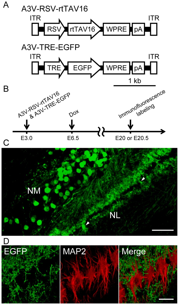
Figure 5. A3V-mediated Tet inducible expression system.
(A) Tet inducible A3V constructs: rtTAV16, reverse tetracycline-controlled transactivator variant 16; TRE, tetracycline response element. (B) A3V-RSV-rtTAV16 and A3V-TRE-EGFP (0.5–1.5 µl, 1×10¹³ GC/ml each) were injected at E3.0, and Dox was administered at E6.5. Immunofluorescence labeling was conducted at E20 (n = 2 embryos) or E20.5 (n = 2 embryos). (C) Anti-EGFP immunofluorescence on a coronal section of the NL-NM circuits at E20.5. The EGFP signal was not detected in the cell bodies of the majority of NL neurons, but strong EGFP signal was observed in the cell bodies of some NL neurons (arrowheads). Scale bar indicates 100 µm. (D) A magnified view of NL neurons at E20.5, visualized with double immunofluorescence labeling for EGFP (green) and dendrite marker MAP2 (red). The immunofluorescence of EGFP was clearly separated from that of MAP2 in the NL dendritic portion. Scale bar indicates 20 µm.

