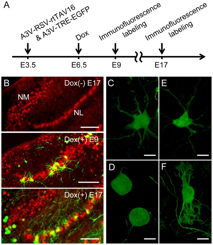
Figure 7. A3V-mediated Tet-inducible system robustly transduced a sparse population of NM and NL neurons.
(A) A3V-TRE-EGFP and A3V-RSV-rtTAV16 (total 0.5–1.5 µl, 5×10¹¹ GC/ml each) were injected at E3.5, and Dox was administered at E6.5. (B) Double immunofluorescence labeling for EGFP (green) and NeuN (red). No apparent EGFP signal was detected at E17 in the Dox (−) preparation (upper), while strong EGFP signal was observed both at E9 (middle) and E17 (bottom) in Dox (+) embryos. Scale bars indicate 100 µm. (C and D) Higher magnification views of EGFP-expressing NM neurons at E9 and E17, respectively. Scale bars indicate 20 µm. (E and F) NL neurons at E9 or E17, respectively. Scale bars indicate 20 µm.

