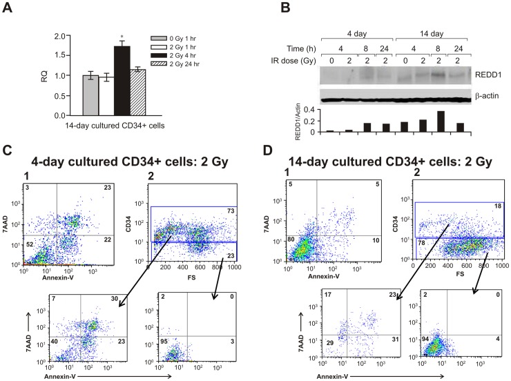
Figure 3. REDD1 expression in human hematopoietic cells.
Purified human CD34+ cells were cultured for 4 and 14 days before being exposed to 2 Gy radiation. (A) mRNA levels for REDD1 in 14-day cultured CD34+ cells using 18S rRNA as a control to calculate the relative quantity (RQ) of gene expression at different times after IR. Means ± SD. *, p <0.05, 2 Gy vs. 0 Gy sham-irradiated control. (B) Western blot shows REDD1 protein levels in cultured CD34+ cells at different times after 2 Gy irradiation. Representative immunoblots and ratio of REDD1/β-actin expression from 3 independent experiments are shown. Flow cytometric analysis for the apoptotic cell death marker Annexin-V/7AAD and CD34 surface marker was performed. Radiation resulted in more apoptotic cells in 4-day (C-1) cultured cells than in 14-day (D-1) culture. (C-2) and (D-2) CD34-positive (progenitor) and CD34-negative (mature) populations were separately gated for Annexin-V/7AAD analysis. FS, forward angle light scatter.

