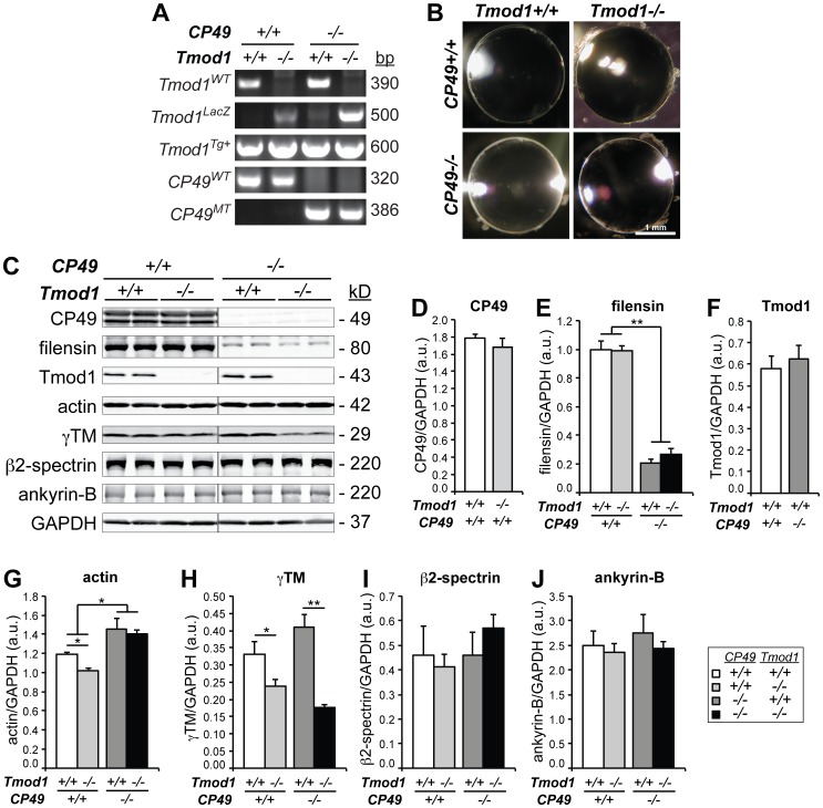
Figure 1. Cytoskeletal protein levels in mouse lenses lacking Tmod1 and/or CP49.
(A) PCR genotyping of Tmod1WT, Tmod1LacZ, Tmod1Tg+, CP49WT, and CP49MT alleles in Tmod1+/+ and Tmod1−/− lenses in both CP49+/+ and CP49−/− backgrounds. (B) Normal anatomy and absence of overt cataracts in 1-4-month-old dissected lenses lacking Tmod1 and/or CP49. (C) Western blots for CP49, filensin, Tmod1, actin, γTM, β2-spectrin, and ankyrin-B in 2-mo-old lenses lacking Tmod1 and/or CP49. GAPDH was used for normalization, with GAPDH probed on each cytoskeletal protein blot. Each lane was loaded with a lens gel sample from an individual mouse. (D-J) Western blot band intensities of (D) CP49, (E) filensin, (F) Tmod1, (G) actin, (H) γTM, (I) β2-spectrin, and (J) ankyrin-B were densitometrically quantified with normalization to GAPDH. Error bars reflect mean±SEM of n = 3 lanes/genotype within a single blot. *, p<0.05; **, p<0.01.

