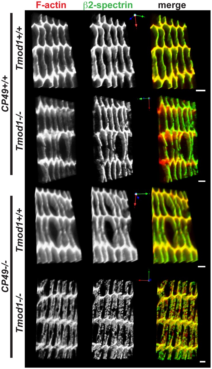
Figure 2. Simultaneous deletion of both Tmod1 and CP49 leads to widespread disruption of F-actin and β2-spectrin organization on lens fiber cell membranes.
Panels depict three-dimensional reconstructions of confocal Z-stacks of equatorial cryosections from 1-mo-old Tmod1+/+ and Tmod1−/− mouse lenses in both CP49+/+ and CP49−/− backgrounds. Cryosections were immunostained for β2-spectrin and phalloidin-stained for F-actin. Spectrin-actin network organization was examined in equatorial sections from 3–4 animals from each genotype, and representative images are shown. Scale bars, 2 µm.

