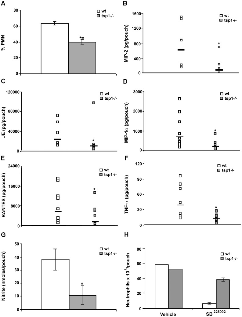
Figure 3. TSP1 increases PMN recruitment to sites of acute inflammation.
Six day-old air pouches received 1 ml of 1% λ-carrageenan, and the mice were sacrificed 24 h after injection. (A) The percentage of infiltrating PMN was quantified in 10 different 400× fields with inflammatory infiltrate. Bars, mean ± SEM, n = 5−6 mice/group. (B–F) The levels of mouse MIP-2, JE, MIP-1α and RANTES, and TNF-α in the air pouch lavage were determined using a MIP-2 Quantikine® ELISA or a multiplexed ELISA array (Quansys Biosciences), as described in Materials and Methods. Data are pooled from two independent experiments and represent geometric mean, n = 5−11 mice/group. (G) 50 µl of exudates were used for NO2 − detection using a Griess Reagent System. All samples were run in triplicate, as described in Materials and Methods. Bars, mean ± SEM, n = 4 mice/group. (H) The absolute number of neutrophils was determined in the presence or absence of SB225002 (50 µM) or vehicle (saline) by FACS, as described in Materials and Methods. Bars, mean ± SEM, n = 3 wt and tsp1−/− mice. *indicates p<0.05; **indicates p<0.001.

