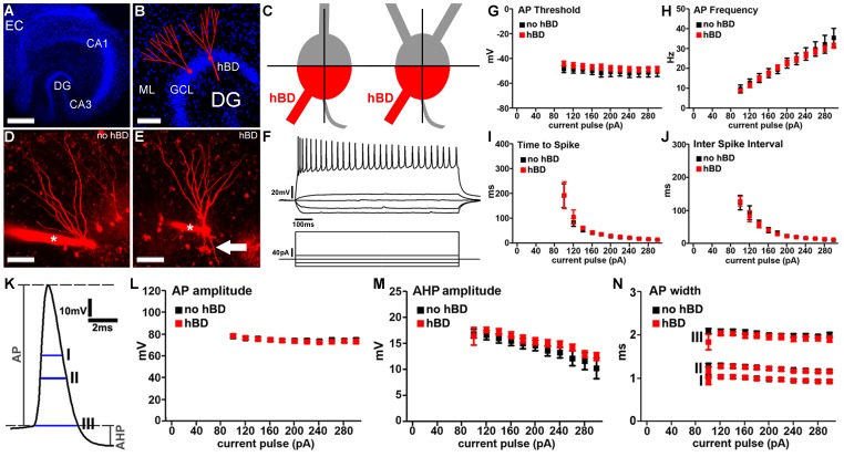
Figure 1. Basic intrinsic cellular properties of dentate granule cells with hilar basal dendrites.
(A) Overview of a mature entorhino-hippocampal slice culture (blue, TOPRO nuclear stain; EC, entorhinal cortex; DG, dentate gyrus; CA1, hippocampal subfield CA1). Scale bar: 500 µm. (B) Dentate gyrus of a mature entorhino-hippocampal slice culture shown at higher magnification. Schematic representations of dentate granule cells. Granule cell somata are located in the granule cell layer (GCL) while granule cell dendrites extend into the molecular layer (ML). A subset of mature dentate granule cells may exhibit additional dendrites, which emerge from the basal portion of the soma and extend into the hilar region (hilar basal dendrites; hBDs; blue, TOPRO nuclear stain). Scale bar: 100 µm. (C) Hilar basal dendrites (hBDs) were defined using the following objective criteria. The granule cell soma was divided into quarters by connecting the origin of the axon with the apical pole of the soma and by an orthogonal centerline trough this line. The two quarters next to the origin of the axon were called basal quarters (red). Dendrites emerging from the basal quarters, reaching into the hilar region and not crossing the centerline were considered as hBDs. (D, E) 2D-projected confocal image stacks of dentate granule cells filled with Alexa568 (red). Asterisk indicates patch electrode. Arrow points to a hBD. Scale bar: 50 µm. (F) Sample traces showing input-output curves of a hBD-GC. Voltage traces (top) in response to a 1 sec current pulse. Protocol is shown underneath the traces. (G–J) Granule cells with and without hBDs were indistinguishable in their (G) actionpotential (AP)-threshold, (H) AP frequency, (I) time to first spike or (J) inter spike interval (n = 9 GCs without hBDs and n = 12 GCs with hBDs; in 7 cultures). (K–N) Evaluation of action potential properties did not show a significant difference between the two GC populations in (L) AP-amplitude, (M) afterhyperpolarization (AHP) amplitude or AP width (N) measured at three different positions (indicated with I, II and III in panel K).

