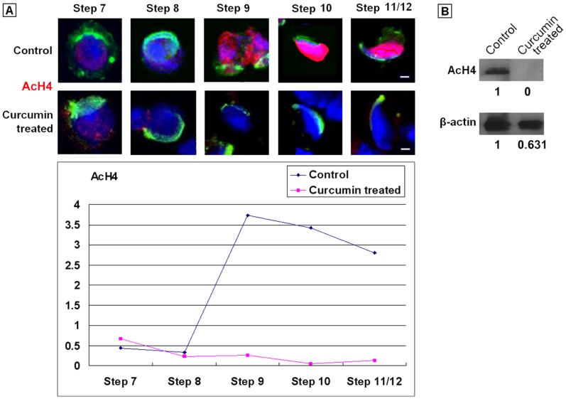
Figure 4. Representative pattern of AcH4 expression in spermatids treated with 50 µM Curcumin for 48.
h. (A). Immunostaining of AcH4 in spermatids. Step: Developmental steps of spermiogensis. Red: Signals of AcH4. Green: Acrosomes highlighted with lectin PNA. Blue: Nuclei counterstained by Hoechst 33342. Bars = 5 µm. The quantitative analysis of Figure 6 was listed in Table S3. (B). Immunoblot of AcH4 in spermatids.

