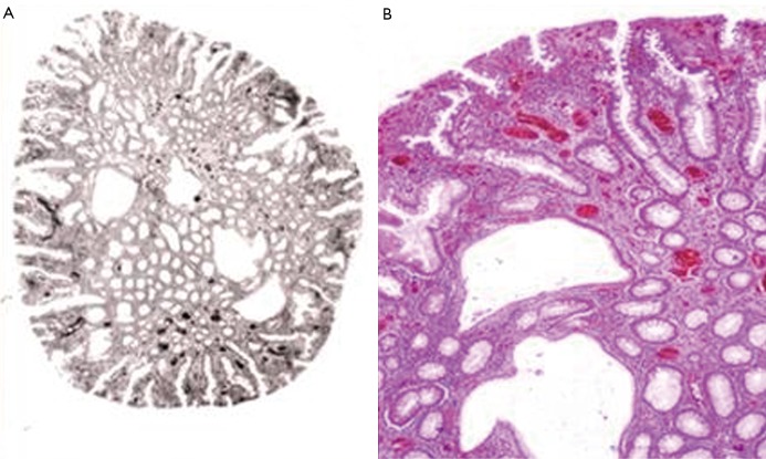
Figure 1.
Hamartomatous polyp from proband’s colon. A. Cystosus glands and regular colonic epithelial glands are visible (organotypic structure) (Hemtaoxilin & eosin (HE) staining). B. Higher magnification of figure 1A (HE staining; 80× magnificiation)

