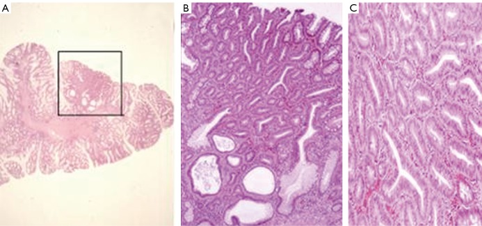
Figure 3.
A. Hyperplastic polyp with adenomatous transformation from proband’s colon. (framed region) (HE staining); B. Adenomatous glands, some of them next to a cystous gland from proband’s colon (HE staining; 80× magnificiation); C. Real neoplastic, tubular adenoma without evidence of malignancy from proband’s colon (HE staining; 80× magnificiation)

