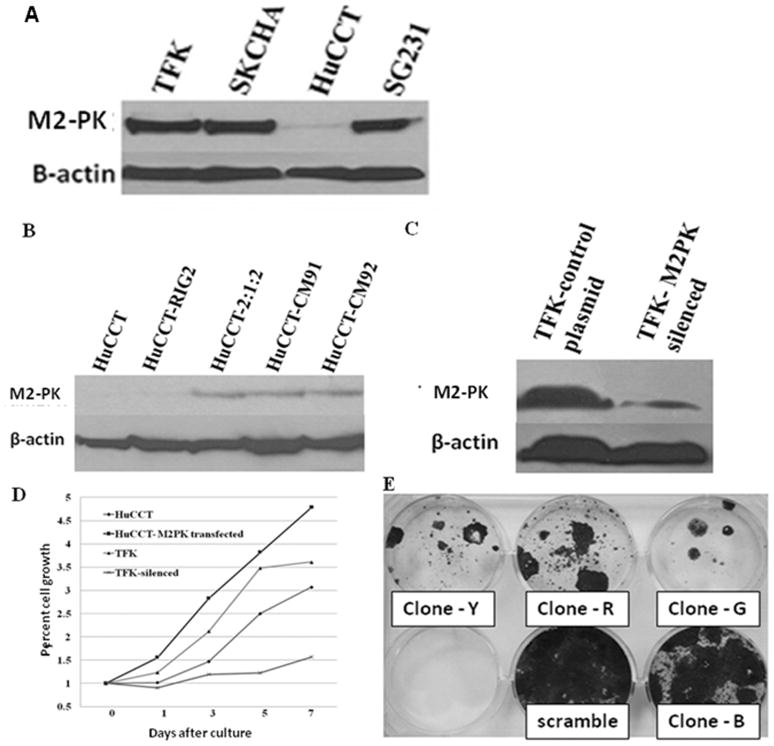
Figure 4.
Strong M2-PK expression was noted in all cell lines except HuCCT(A). M2-PK western blot of HuCCT following transfection with (right 3 lanes) or without (2nd lane) full length M2-PK (B). M2-PK western blot of mock-treated TFK cells and TFK cells silenced with shRNAs (clones G, R and Y) (C). Cell proliferation rates of mock-treated HuCCT, M2-PK transfected HuCCT, mock-treated TFK and M2-PK silenced TFK cells (D). MTS assay showed increased cell proliferation in M2-PK transfected HuCCT cells and significant growth inhibition in M2-PK silenced cells (D). Anti-proliferative effect of different shRNA clones are shown; all clones except clone B had significant growth inhibition (E).

