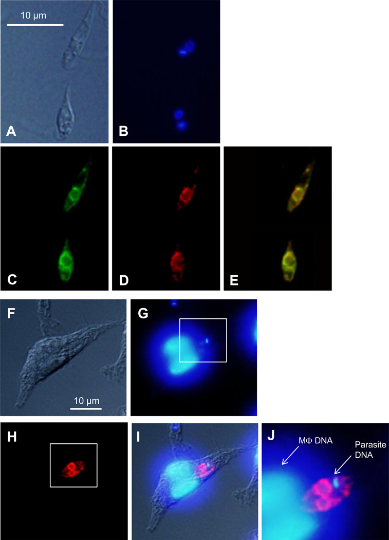
Figure 6. Localization of PLA2/PAF-AH in L. major.
(A–E) Log phase promastigotes of pla2−/+PLA2-GFP were subjected to immunofluorescence microscopy as described in Materials and Methods (2.4). (A) DIC image. (B) DNA staining with Hoechst 33342. (C) PLA2-GFP epifluorescence. (D) Immuno-staining with anti-BIP antibody to label ER. (E) Merge of C and D. (F–J) BM-MΦs were infected with pla2−/+PLA2-GFP parasites for 48 hours and subjected to immunofluorescence microscopy as described in Materials and Methods (2.4). (F) DIC image of an infected MΦ. (G) DNA staining with Hoescht 33342. (H) Immuno-staining with rabbit anti- GFP serum followed by goat anti-rabbit IgG conjugated with Texas Red. (I) Merge of F, G, and H. (J) Merged and magnified image of the boxed areas in G and H.

