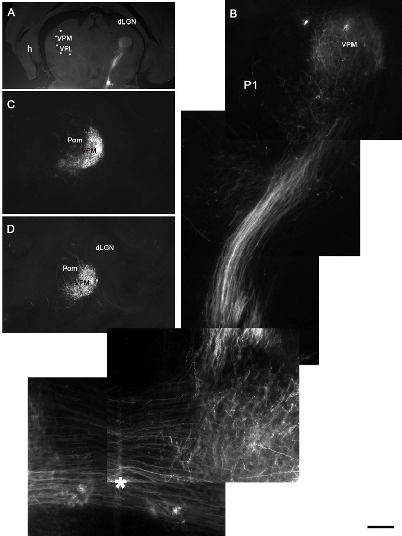
Figure 5.
Postnatal development of the trigeminal lemniscal pathway. A, B: Oblique horizontal sections show that the trigeminal lemniscus has invaded the VPM and axons branch diffusely across the entire nucleus at P1. C, D: Consecutive coronal sections showing the confinement of PrV axons to the VPM, with only a few axons spilling into the neighboring posteromedial nucleus (Pom). Note that no lemniscal axons enter the dorsal lateral geniculate nucleus (dLGN). Asterisk in B indicates the midline crossing of the PrV axons. Scale bar = 1200µm for A, 200µm for B and 500µm for C, D.

