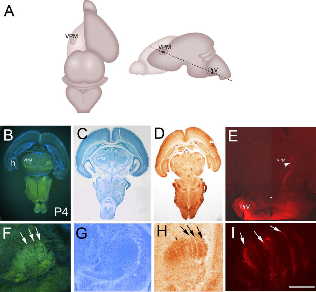
Figure 7.
A: Schematic illustration of the oblique horizontal plane of sectioning to preserve the PrV-thalamic pathway. B: VGLUT2 immunostained oblique horizontal section with DAPI counterstain from a P4 brain. C: Nissl stain. D: Cytochrome oxidase histochemistry. E: DiI labeling at P4. H: hippocampus; VPM: ventroposteromedial nucleus. F–I show higher magnification views of the VPM with VGLUT2 immunohistochemistry (F), Nissl (G), CO (H) staining and DiI (I) labeling. Barreloid rows are clearly visible with VGLUT2 immuno- and CO histochemistry (arrows). Note also alignment of axon terminals along bands (arrows in I), reflecting the barreloid organization in the VPM. Scale bar in F = 200µm, I = 200µm.

