Fig. 1.
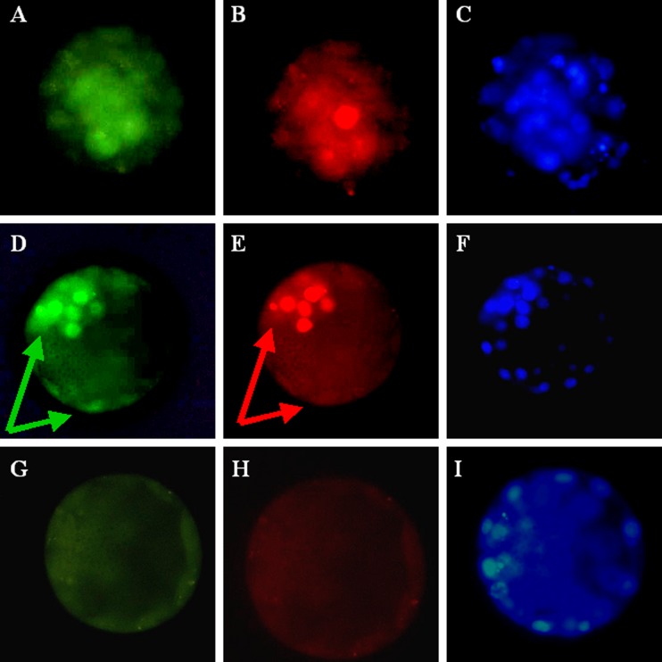
Representative pictures show double staining of OCT4 and NANOG in the human morula and blastocyst. a Immunostaining of OCT4 in the morula. b Immunostaining of NANOG in the same morula. c DAPI staining in the morula. d Staining of OCT4 in the blastocyst. Immunostaining is seen both in the inner cell mass and the trophoblast; arrows. e Staining of NANOG in the same blastocyst. Staining is seen only in the inner cell mass; arrow. f DAPI staining in the same blastocyst. g Exclusion of OCT4 antibody. h Exclusion of NANOG antibody. i DAPI staining of the embryo without primary antibodies present

