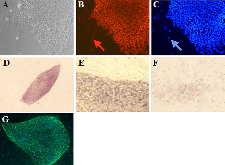
Fig. 3.
Immunostaining of OCT4 and NANOG in human embryonic stem cells, and in situ hybridization of NANOG in embryonic stem cells. a Light microscopy of human embryonic stem cells. b NANOG immunostaining in embryonic stem cells. The feeder cells do not show staining for NANOG (red arrow). c Nuclear staining of embryonic stem cells and fibroblast cells. DAPI staining is seen in feeder cells (blue arrow). d & e In situ hybridization showing NANOG mRNA in embryonic stem cells. NANOG f Mouse embryonic stem cells hybridized with human NANOG primer. g OCT4 immunostaining of stem cell line HS426 (green). Blue colour shows nuclear DAPI staining

