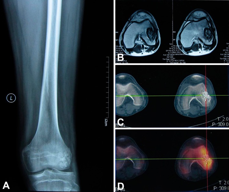
Fig. 1A–D.
The images illustrate the case of a 24-year-old man with a diagnosis of recurrent Ewing’s sarcoma who received intralesional curettage and autogenous cancellous bone graft in his hometown hospital because of misdiagnosis. (A) A preoperative AP radiograph shows the extension of the tumor that compromised the lateral femoral condyle. (B) The MRI scans determined tumor extension. (C) The CT scan showed the area of bone destruction. (D) The bone scan showed increased uptake in the area of tumor invasion. These images were used to design the levels of tumor resection with the computer-assisted navigation system.

