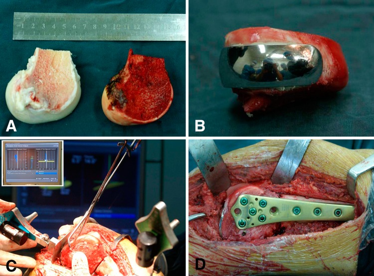
Fig. 3A–D.
The photographs show the unicondylar osteoallograft prosthesis composite reconstruction that was performed using computer-assisted navigation. (A) After thawed, the osteoarticular allograft was cut into the proper size to match the bone defect The allograft is on the left and the resected femoral condyle is on the right. (B) The cemented femoral component was fixed to the allograft to form a unicondylar osteoallograft prosthesis composite. (C) The navigation system enabled the surgeons to precisely implant the unicondylar osteoallograft prosthesis composite. Congruency of the articular surface was meticulously adjusted. The insert in the upper left corner shows mechanical alignment was checked to avoid a varus or valgus deformity. (D) The unicondylar osteoallograft prosthesis composite was fixed to the host bone with a locking plate.

