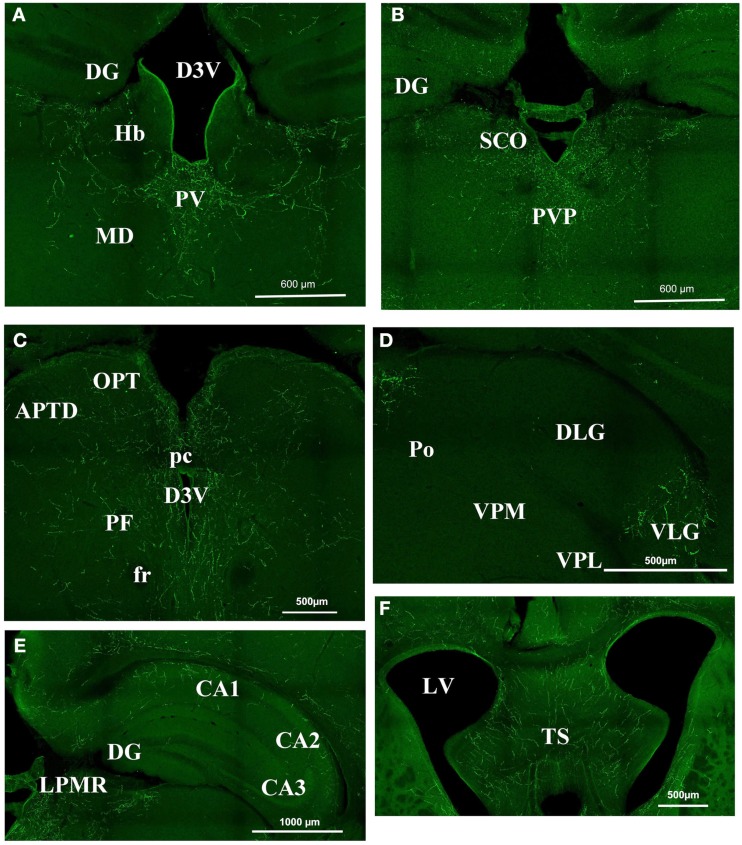
Figure 2.
Orexin staining in the thalamus, hippocampus, and septum. (A) Periventricular area Bregma −1.82. Strong staining is visible at the level of the PV, while there is no staining in the DG, the Hb, and the MD. (B) Periventricular area at Bregma −2.30. Note the strong staining in the PVP and SCO and still the absence of staining in the DG. (C) Periventricular area at Bregma −2.80. There is strong staining around the third ventricle, but staining is faint in the OPT and the APTD. (D) Geniculate nucleus. Orexin is present in the VLG but absentin the DLG, the Po, the VPM, and VPL. (E) Hippocampal structures at Bregma −2.3. Note the staining in CA1 CA2 but not in CA3. (F) Strong staining in the TS. APTD, anterior pretectal nucleus dors.; D3V, dorsal third ventricle; DG, dentate gyrus; DLG, dorsal lateral geniculate; fr, fasciculus retroflexus; Hb, habenular nucleus; LPMR, lat. post. thal. nu. LV, lateral ventricle; MD, medio dorsal thalamus nucleus; OPT, olivary pretectal nucleus; pc, post commissure; PF, para fascicular thalamus nucleus; Po, post. thal. nu.; PV, paraventricular thalamic nucleus; PVP, paraventricular thalamic nucleus post; SCO, sub commissural organ; TS, triangular septal nucleus; VLG, ventral lateral geniculate; VPL, ventro postero lateral thal; VPM, ventro postero med. thal.

