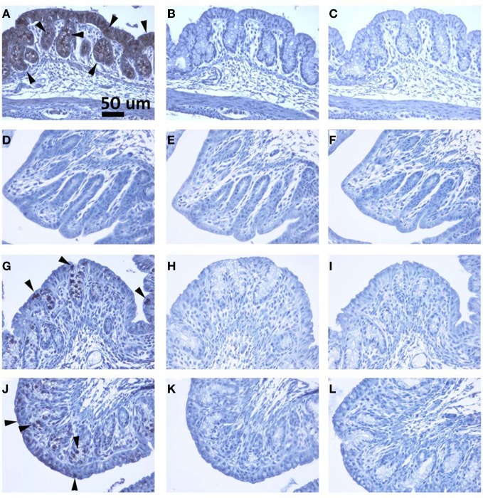
Figure 6.
(A–C), (D–F), (G–I) and (J–L) are colon sections from naïve, Stx2, heat-inactivated Stx2 and Stx2 mutant treated rabbits, respectively. (A, D, G and J) are sections stained with anti-Gb3 antibody. Strong signal in A (luminal layer) and less intense stain in G and J can be recognized, whereas no signal is detected in D (Stx2 treated). Arrowheads show examples of Gb3 positive spots. In (B, E, H and K), isotype matched rat IgM was used in the place of anti-Gb3 antibody to serve as a negative control. (C, F, I and L) are primary antibody omitted, buffer control (negative control). All controls shown are negative. A scale bar in (A) indicates 50 mm.

