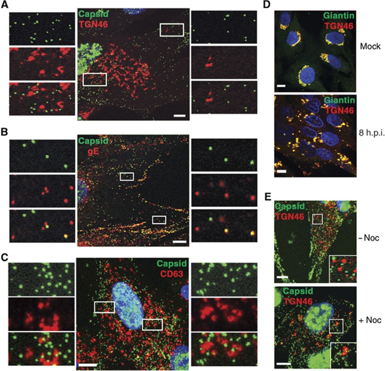
Figure 3.
HSV1 capsids colocalise with glycoprotein E but not TGN46 or CD63 in HFFF-2 cells. Cells grown on coverslips were infected with the GFP-VP26 virus (green) (A–C, E) or s17 (D) at a multiplicity of 2, and fixed 8 h after infection. Cells were stained with antibody against (A, E) TGN46 (red), (B) gE (red), (C) CD63 (red) or (D) TGN46 (red) and giantin (green), and nuclei stained with DAPI (blue) before imaging with a Zeiss LSM510 Meta confocal microscope. In (E), nocodazole was added at a concentration of 2 μg/ml for 30 min before fixing to depolymerise the microtubule network (+noc). Scale bar=10 μm.

