Figure 1.
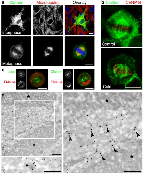
Clathrin was targeted to the mitotic spindle of NRK cells. a, Confocal micrographs showing the subcellular distribution of clathrin at interphase and metaphase. GFP-LCa (left, green), α-tubulin (centre, red) and nucleic acids (blue). b, Cells expressing GFP-LCa fixed before (left) or after (right) depolymerisation of non-kinetochore microtubules. c-d, The association of clathrin with microtubules was not via coated membranes. c, Example images of live cells expressing either GFP-α-tubulin (left) or GFP-LCa (right) imaged following 24-28 h incubation with FM4-64 (red). d, Association of clathrin with microtubules visualised by immunogold EM. CHC (15 nm) and α-tubulin (10 nm gold) in mitotic NRK cells. Chromosomes are denoted by asterisks. A morphologically distinct CCV (ii) is indicated by an arrow. Arrowheads denote CHC labelling associated with microtubules. Scale bars, 10 μm (a-c) 250 nm (d).

