Figure 2.

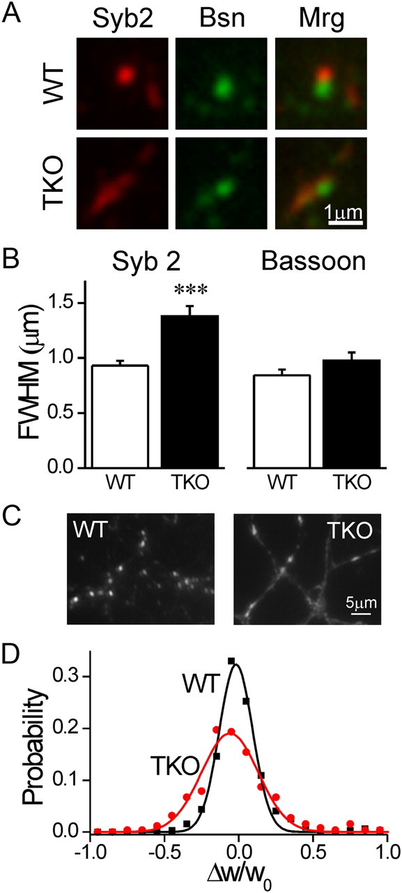
Synaptic vesicle clusters are dispersed in synapsin TKO neurons. A, Immunostaining forSyb2 (red) and Bassoon (Bsn, green) in WT (top) and synapsin TKO (bottom) neurons. Merge (Mrg) images appear at right. Bassoon staining, representing the active zone, appears similar in both genotypes, while Syb2 staining, representing the vesicle cluster, is spread out. B, Average FWHM width of Syb2 (left) and Bassoon (right) puncta (mean ± SEM). C, Representative images of SypI-EGFP puncta in WT (left) and synapsin TKO (right) neurons. Notice that in TKO neurons SypI-EGPF is distributed along longer segments of the axon and that the axon is more readily seen. D, Fractional change in the width of puncta over a period of 10 min. The distribution of Δw/w0 values, representing the change in synaptic width, indicates that while synapses of both genotypes tended to maintain their dimensions, the variability in TKO puncta was larger, even considering that they were wider to begin with. ***p < 0.001, Student's t test.
