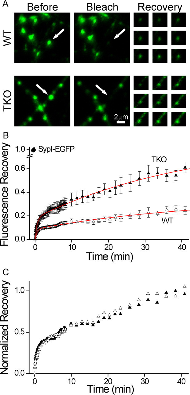
Figure 4.

The fraction of mobile synaptic vesicles is determined by the synapsins. Mobility of synaptic vesicles was probed by FRAP of SypI-EGFP. A, Representative FRAP experiments in WT (top) and synapsin TKO (bottom) neurons. Shown is a field including several synaptic puncta before (left) and immediately after (middle) bleaching of a single punctum (white arrow). The fluorescence of the affected synapse is reduced to >85% of its original value, while neighboring synapses are unaffected. The recovery of fluorescence is shown at right 1, 6, 11.5, 16, 20.5, 26, 31, 36, and 41 min after bleaching, with the contrast homogeneously increased to improve visibility. Recovery in the synapsin TKO neuron is noticeably faster. B, Average time course of FRAP experiments in WT (open triangles) and TKO (solid triangles) synapses (mean ± SEM). Intensity data are normalized by the initial fluorescence (leftmost symbols), and lowest value is shifted to 0. Sampling was performed at intervals of 5, 15, 90, and 150 s. The data are corrected by changes in the intensity of unbleached synapses in distant areas of the image. Red lines represent global fitting of the data with a double exponential function, where the time constants are shared. Recovery in TKO neurons is more extensive. C, Same data as in B, normalized by the average of the three end points. Recovery proceeds with equal kinetics, as evidenced also by the global fit lines in B.
