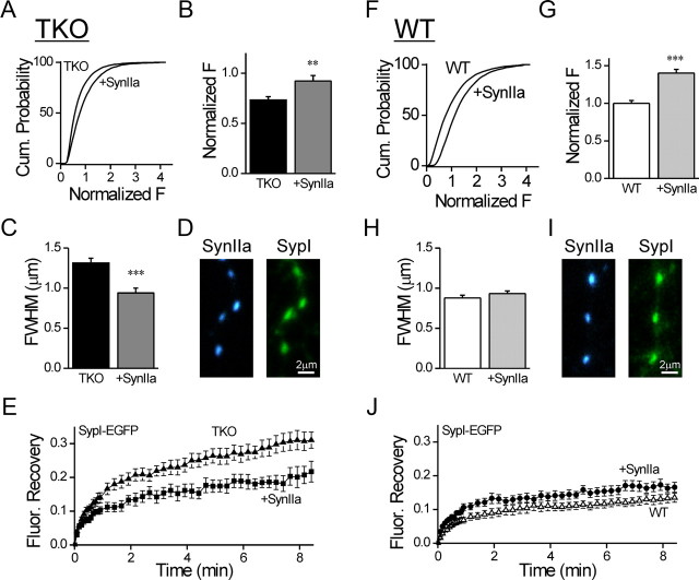
Figure 9.
Synapsin IIa rescues the immobilization and clustering of vesicles in synapsin TKO neurons. A, Cumulative plot of Syb2 immunofluorescence in synaptic puncta of TKO neurons expressing EGPF-synapsin IIa compared with uninfected neurons. B, Average of the means of Syb2 signal calculated for independent images (mean ± SEM). EGFP-synapsin IIa increases the intensity of Syb2 staining. G, The data were normalized by the mean intensity observed in synapses of WT neurons. C, Average FWHM width of the Syb2 signal in synaptic puncta in TKO neurons expressing EGFP-synapsin IIa compared with uninfected neurons. D, Representative images of synapses in TKO neurons coexpressing TagBFP-Synapsin IIa (blue, left) and SypI-EGFP (green, right). E, Effect of coexpression of TagBFP-Synapsin IIa (filled squares) on the FRAP time course of SypI-EGFP in TKO neurons (filled triangles). TagBFP-Synapsin IIa reduced the mobility of the vesicles. F–J is the same as A–E except performed in WT neurons. Synapsin IIa increased the intensity of Syb2 puncta in WT neurons, but did not affect their width, or the time course of FRAP. **p < 0.01, ***p < 0.001.

