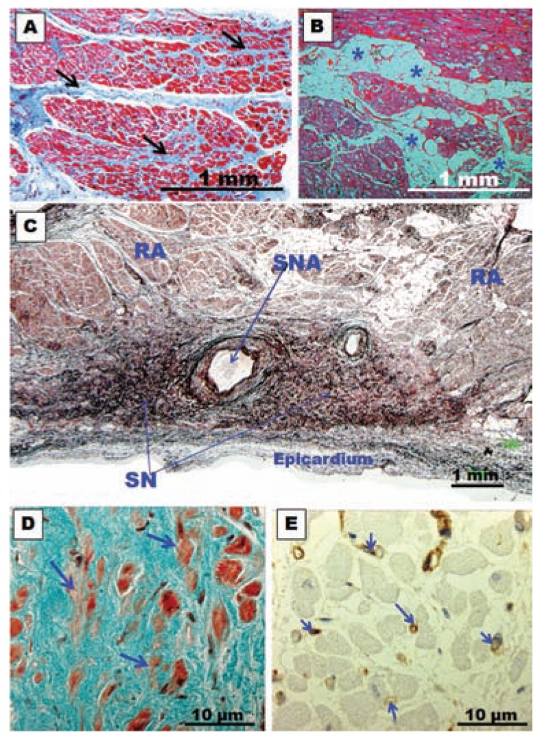
Fig. (12).
(A) and (B)Histological cross-section biopsy samples from the posterior left atrial wall in specimens with chronic atrial fibrillation for rheumatic mitral valve disease. Note in (A) the abundant connective tissue between the myocytes (arrows) and interstitial fibrosis. In (B) note accumulation of fat cells (asterisks) between myocytes. (C) Histological section of the sinus node with methenamine silver staining of a specimen with chronic atrial fibrillation. Note the intense accumulation of connective tissue between nodal cells. SN, sinus node; SNA, sinus node artery; RA, right atrium. (D) Sinus node. Trichrome stain. Note the abundant connective tissue and fewer larger myocardial nodal cells (arrows) in a patient with long-term chronic atrial fibrillation. (E) Immunohistochemical staining for CD31 (vessel walls stained in brown) of the sinus node in a patient with long-term chronic atrial fibrillation. Note fewer and thinner capillaries in the sinus node (arrows).

