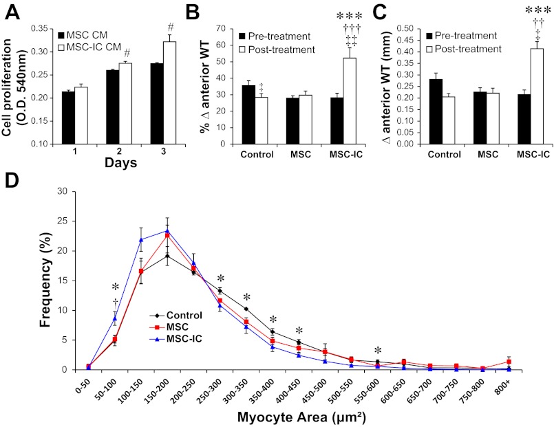
Fig. 10.
Poly(I:C)-treated MSC effectively promotes formation of new cardiomyocytes. A: HL-1 cardiomyocytes preplated in a 24-well plate were treated with 50% MSC-conditioned medium (MSC CM or MSC-IC CM). Cell proliferation was monitored by MTT assays for 3 days, comparing the difference between MSC CM and MSC-IC CM at the time points indicated; n = 3. B and C: echocardiography was used to measure % anterior wall thickening (B) and anterior wall thickness (Δanterior WT) (C). Heart tissue sections were prepared 1 mo after the low-dose (1 × 10⁶ cells/kg) MSC therapy. H&E-stained paraffin sections were used for morphometric analysis. D: myocyte size distribution analysis derived from the morphometric analysis. n = 4 per group; *P < 0.05 and ***P < 0.001 saline control vs. MSC-IC; †P < 0.01 and †††P < 0.001 untreated MSC vs. MSC-IC; ††P < 0.01 untreated MSC vs. MSC-IC; ‡P < 0.05; ‡‡P < 0.01 vs. pretreatment; #P < 0.01 vs. MSC CM.

