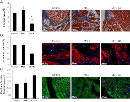
Fig. 9.
Antifibrotic, antiapoptotic, and proangiogenic activities are promoted by poly(I:C)-treated MSC. Heart tissue sections were prepared 1 mo after the low-dose (1 × 10⁶ cells/kg) MSC therapy and processed for analysis of fibrosis, apoptosis, and angiogenesis. A: analysis of myocardial fibrosis by trichrome staining. B: analysis of myocardial apoptosis by TUNEL staining. Myocytes were stained by a troponin T antibody (red). Total nuclei were stained by DAPI (blue). Apoptotic nuclei are cyan colored. C: analysis of capillary density by GSL-IB4 staining. Myocytes were stained by a troponin T antibody (green). Total nuclei were stained by DAPI (blue). Capillaries are red/pink colored. Representative images of fibrosis, apoptosis, and stained capillaries for each group are illustrated (×200). n = 3–4 per group; *P < 0.05 vs. saline control; †P < 0.05 and ††P < 0.01 vs. untreated MSC.

