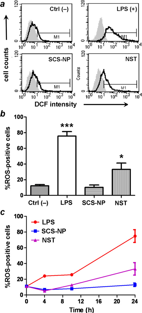
Fig. 7.
(a) Flow cytometry of ROS production in RAW cells exposed to various stimuli (t = 24 h): control (upper left), LPS (upper right), SCS-NP (lower left), and NST (lower right). (b) ROS production is stimulated strongly by LPS (p < 0.001, ***) and by NSTs (p < 0.05, *), but not by SCS-NPs. (c) ROS production in stimulated macrophage as a function of incubation time. All experiments were performed in triplicate.

