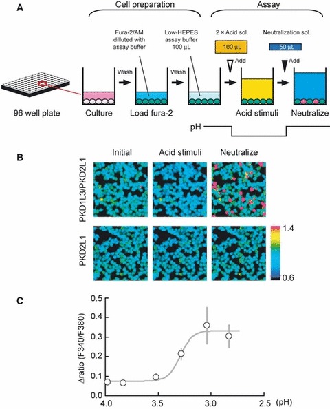
Fig 1.

The neutralization method, a novel method for evaluating PKD1L3/PKD2L1 activity. A novel Ca2+ imaging method using neutralization was developed. (A) Schematic representation of the method. The cell preparation and assay steps are illustrated. In the assay step, cells are exposed to acid for 8 s and then neutralized. The lower black line schematically indicates the pH change of the extracellular solution. (B) Representative ratiometric images of Fura-2 loaded cells at the initiation of the assay, during acid stimulus and after neutralization. Upper panels indicate cells transfected with PKD1L3 and PKD2L1. Lower panels indicate cells transfected with only PKD2L1. The color scale indicates the F340/F380 ratio. Scale bar, 100 μm. (C) The pH-dependent cell response was evaluated using the neutralization method. Cells were transfected with PKD1L3 and PKD2L1. Each point represents the mean ± SEM of the fluorescence ratio change in independent trials (n = 6). In each trial, 100 DsRed2-positive cells were analyzed.
