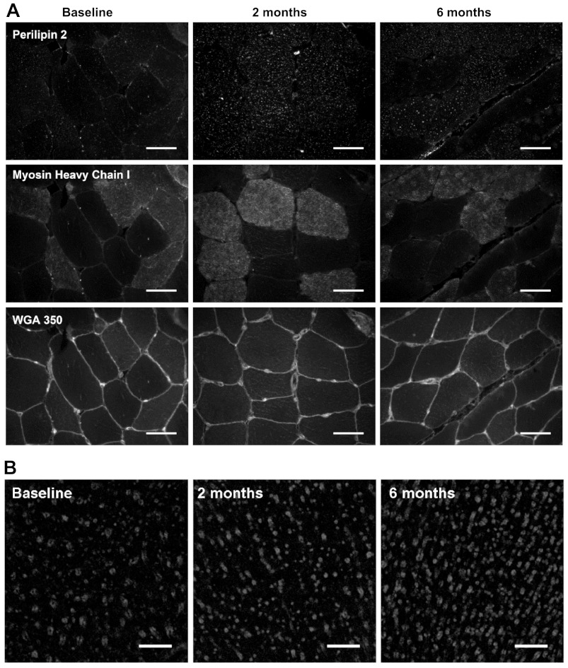
Fig. 3.
Immunofluorescence images of fiber type-specific perilipin 2 protein expression. A: representative images of perilipin 2 at baseline and 6 mo, stained using anti-perilipin 2 in combination with anti-myosin heavy chain type I and wheat germ agglutinin 350 (WGA350) and viewed and quantified with wide-field immunofluorescence microscopy. Scale bar, 50 μm. B: representative confocal images of perilipin 2 at baseline and 6 mo; rings of perilipin staining are clearly visible in both images. Scale bar, 10 μm.

