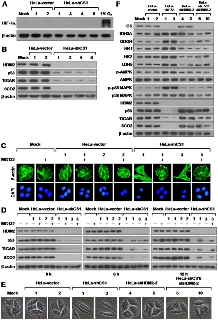
Figure 6. CS knockdown induced EMT switch is reverted by p53 reactivation.
(A) Western blotting of HIF-1 α expression in CS knockdown cells. Total proteins isolated from cells as indicated were blotted with antibodies for HIF-1α and β-actin. Proteins prepared from cells cultured in 1% O2 for 24 h serves as positive control for hypoxic condition. (B) Western blotting of HDM2, p53, TIGAR, and SCO2 expression in CS knockdown cells. Total proteins isolated from indicated cells were blotted with antibodies as labeled. (C) Fluorescence imaging of stress fibers of CS knockdown cells treated with MG132. Cells were treated with 10 mM MG132 for 12 h and then stained with Alexa Fluor 488-conjugated phalloidin and DAPI. (D) Western blotting of HDM2, p53, TIGAR, and SCO2 proteins in CS knockdown cells treated with MG132. Cells as indicated were treated with 10 mM MG132 for 0, 6, and 12 h. Total protein extracts isolated from these cells were blotted with antibodies as indicated. (E) Morphological imaging of CS, HDM2, and CS/HDM2 knockdown cells. Cells with single CS, HDM2, or double CS/HDM2 knockdown as indicated were selected and imaged. (F) Western blotting of proteins involved in bioenergetic metabolism in single CS, HDM2, and double CS/HDM2 knockdown cells. Total protein extracts isolated from the indicated cells were blotted with antibodies as labeled. The level of β-actin serves as a loading control.

