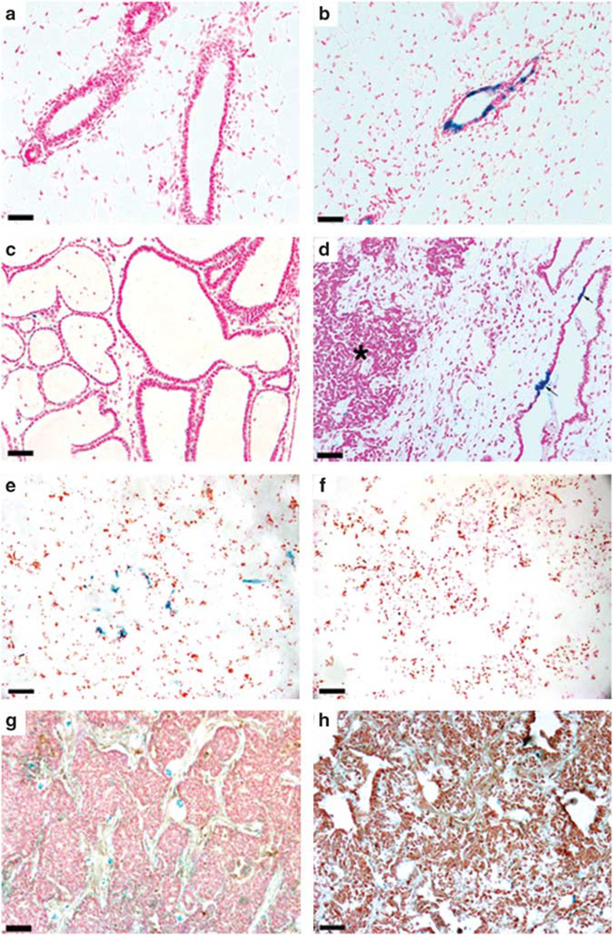
Figure 1.
PI-MECs are present in WAP-Int3/WC/R26 mammary glands but do not form tumors. Nulliparous (a) and primiparous (b) WC/R26 control glands demonstrate the marking of PI-MECs with β Gal following pregnancy and involution. (c) A cross-section of a 3-day post-partum WAP-Int3/WC/R26 mammary gland demonstrating lack of secretory lobular development and the presence of cystic ducts. Note structures lack any milk fat in their lumens. (d) Following pregnancy and involution, β Gal+ cells were found in normal mammary epithelium (arrows) but not in tumors (asterisk). (e) Cultures of mammary epithelial cells taken from parous non-pregnant WAP-Int3/WC/R26 mice contain a small population (6%) of β Gal+ cells (blue). (e) Cultures of mammary tumors taken from parous non-pregnant WAP-Int3/WC/R26 mice contain no β Gal+ cells. (g, h) WAP-Int3/WC/R26 tumors stained negative for Cre expression during pregnancy (g) and positive for Neo expression following pregnancy (h). Scale Bars: a, b, e and f = 100 µm; c, d = 250 µm.

