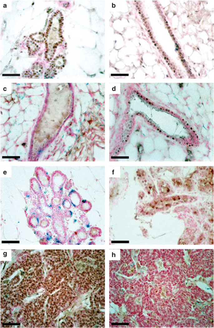
Figure 2.
WAP-Int3/WC/R26 mice do not maintain normal PR+ or RANKL+ epithelial cell populations. ERα staining in WAP-Int3/WC/R26 glands (a) and normal glands (b) demonstrates no significant difference between the percentage of ERα+ epithelial cells in transgenic and control glands (30.4 and 23.1%, respectively). PR staining in WAP-Int3/WC/R26 glands (c) and normal mammary glands (d) demonstrates significantly lower percentage of PR+ epithelial cells in transgenic glands (0.16 and 14.0%, respectively; P < 0.05). RANKL staining in staining in WAP-Int3/WC/R26 glands (e) and normal mammary glands (f) demonstrates the transgenic glands also have a significantly lower percentage of RANKL+ cells (0.43 and 13.4%, respectively; P < 0.05). Tumors from WAP-Int3/WC/R26 mice are ERα+ (g) and PR− (h). Scale Bars = 100 µm.

