Figure 3.
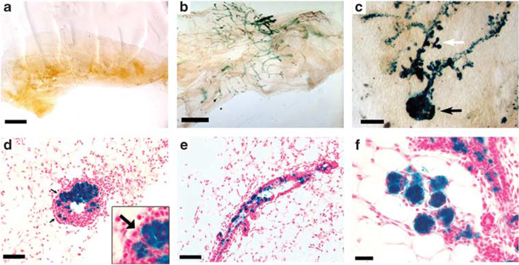
Dissociated WAP-Int3/WC/R26 PI-MECs contribute to mammary gland regeneration when transplanted with normal MECs. (a) A total of 2 × 10⁶ dissociated WAP-Int3/WC/R26 MECs failed to regenerate a gland upon transplantation into a cleared mammary fat pad of a nu/nu female host. (b) When 2 × 10⁵ dissociated WAP-Int3/WC/R26 MECs were mixed with 1 × 10⁵ or 2 × 10⁵ normal MECs, PI-MECs (blue cells) contributed to the resulting complete outgrowths. (c) Higher magnification image of a whole-mounted mammary gland taken from a 8-day pregnant mouse 22 days following inoculation with a mix of 2 × 10⁵ WAP-Int3/WC/R26 and 2 × 10⁵ normal MECs demonstrating contribution of WAP-Int3/WC/R26 PI-MECs to both end buds (black arrow) and developing side branches (white arrow). (d) Cross-section of an end bud in a developing WAP-Int3/WC/R26 and normal MECs chimeric gland demonstrating contribution of the WAP-Int3/WC/R26 PI-MECs to the body of the end bud, but not the cap cells (arrows). Inset is a higher magnification of the cap cells of the same image. (e) Cross section of a duct from a chimeric gland demonstrating presence of WAP-Int3/WC/R26 PI-MECs’ progeny. (f) Cross-section of and 8-day pregnant chimeric gland demonstrating contribution of WAP-Int3/WC/R26 PI-MECs to the developing acinar structures. Scale Bars: a and b = 1000 µm; c = 200 µm; d and e = 150 µm; f = 50 µm.

