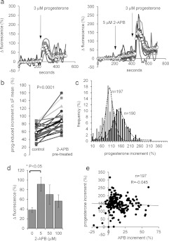
Figure 3. 2-APB modulates the progesterone-induced [Ca2+]i transient.
(a) Elevation of [Ca2+]i at the PHN in response to stimulation with 3 μM progesterone (arrow) under control conditions (left-hand panel) and after 200 s exposure to 5 μM 2-APB (right-hand panel). Both experiments used cells from the same preparation. Traces show 6–8 representative single-cell responses and ΔFmean (○-○) for all 107 (left-hand panel) and 125 (right-hand panel) cells in the experiment. (b) Summary of results from 20 pairs of control and 5 μM 2-APB-pre-treated experiments. Each point shows the mean amplitude of the progesterone-induced transient (increment in ΔFmean) for all of the cells in a single experiment (50–200 cells). Joined pairs of points show 5 μM 2-APB pre-treatment (right-hand point) and corresponding control (left-hand point) using cells from the same ejaculate – such as the pair shown in (a). Data from 20 pairs of experiments are shown and the overall mean for all 20 is shown by Δ-Δ. (c) Effect of 5 μM 2-APB on amplitude distribution of single-cell progesterone-induced transients. Grey bars show the control, black bars show the parallel 5 μM 2-APB-pre-treated experiment. (d) Dose-dependence of potentiation by 2-APB of the progesterone-induced [Ca2+]i transient. Each bar shows the mean amplitude±S.E.M. for four sets of experiments (50–200 cells each). In each set, four experiments were carried out with samples from the same ejaculate, using 0, 5, 50 or 100 μM 2-APB applied 200 s before progesterone. Only 5 μM 2-APB significantly enhanced the [Ca2+]i transient. (e) Amplitude of 2-APB-induced resting [Ca2+]i elevation (APB increment; x-axis) is not correlated with the amplitude of subsequent progesterone-induced [Ca2+]i transient (progesterone increment; y-axis). Results are from 197 cells in one experiment.

