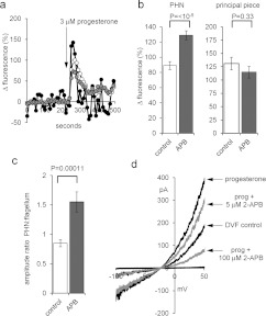
Figure 4. 2-APB does not enhance the flagellar Ca2+ signal or potentiate activation of CatSper by progesterone.
(a) [Ca2+]i (OGB) signal from the PHN (white circles), midpiece (grey circles) and flagellum (black circles) in response to application of 3 μM progesterone (arrow). Each trace shows the mean response from the same nine cells. (b) Amplitude of progesterone-induced [Ca2+]i transient at the PHN (left-hand panel) and midpiece (right-hand panel) under control conditions (white bars; n=43 cells) and after pre-treatment with 5 μM 2-APB (grey bars; n=57 cells). (c) Ratio of [Ca2+]i transient amplitudes simultaneously recorded from the PHN and flagellum under control conditions (white bar; n=43) and after pre-treatment with 5 μM 2-APB (grey bar; n=57). (d) Monovalent currents (DVF control) were enhanced by 500 nM progesterone (upper black trace). Subsequent application of 5 μM 2-APB (upper grey trace) and 100 mM 2-APB (lower grey trace) reduced the amplitude of outward and inward currents.

