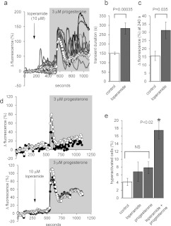
Figure 8. Loperamide potentiates the response of human sperm to progesterone.
(a) Pre-treatment with 10 μM loperamide (arrow) followed by application of 3 μM progesterone (shading). Traces show nine representative single-cell PHN responses and ΔFmean (○-○) for all 81 cells in the experiment. Loperamide elevates resting [Ca2+]i and subsequent exposure to progesterone induced an initial [Ca2+]i transient followed by large [Ca2+]i oscillations. (b) Duration of the progesterone-induced [Ca2+]i transient in the PHN was increased by loperamide pre-treatment. Bars show means±S.E.M. for 11 paired experiments. (c) Progesterone-induced sustained [Ca2+]i increase (ΔFmean at 240 s after progesterone) was enhanced by loperamide pre-treatment. Bars show means±S.E.M. for nine paired experiments. (d) Mean normalized fluorescence (ΔFmean) in the PHN (○-○) and in the midpiece (●-●) under control conditions (upper panel; mean of 19 cells) and after pre-treatment with 10 μM loperamide (lower panel; mean of 33 cells). Potentiation by loperamide of [Ca2+]i transient duration and sustained [Ca2+]i elevation are similar in the two compartments. (e) Loperamide enhances progesterone-induced hyperactivation. Each bar shows the percentage of hyperactivated cells (means±S.E.M.; n=7). Progesterone (3 μM) and loperamide (10 μM), applied individually, failed significantly to increase hyperactivation (not significant; NS). When cells were pre-treated with loperamide (3 min), progesterone significantly increased the proportion of hyperactivated cells over all the other conditions (*P<0.02).

