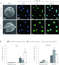
Figure 2. Degranulation of pig neutrophils by the calcium ionophore A23187.
SEM and confocal microscopy of quiescent and activated porcine neutrophils (A) showing DNA (blue) and Pr3 (green), chosen here as a representative NSP. Proteolytic activities as measured by the increase in fluorescence units/s of FRET substrates specific for each NSP in suspensions of 1.5×10⁶ /150 μl of pig neutrophils (median±interquartiles, n=10) and human neutrophils (median±interquartiles, n=5) (B). * indicate significant (α=5%) increases over unstimulated cells. FRET substrates were Abz-QPMAVVQSVPQ-EDDnp for NE, Abz-TPFSGQ-EDDnp for cat G and Abz-VADCADYQ-EDDnp for Pr3 [24–26].

